Retinal Nerve Cells Grown In Lab Could Help Blind
So, you just read an article heading that may surprise you. Yes, scientists have made it possible to grow retinal nerve cells in lab.
Scientists developed a method to turn human stem cells into retinal nerve cells that transmit visual signals from the eye to the brain. Study leader Donald Zack, from the Johns Hopkins University School of Medicine in US, said the study would not only give a better insight of the biology of the optic nerve, but with that it will also provide a cell-based human model that could be used to discover drugs that stop or treat blinding conditions. Adding more, he said that this could lead to the development of cell transplant therapies that restore vision in patients with glaucoma and MS.
Retinal Nerve Cells Grown
The laboratory process, described in the journal Scientific Reports, states
Genetically modifying a line of human embryonic stem cells to become fluorescent upon their differentiation to retinal ganglion cells, and then using that cell line for development of new differentiation methods and characterization of the resulting cells. Using a genome editing laboratory tool called CRISPR-Cas9, investigators inserted a fluorescent protein gene into the stem cells’ DNA. This red fluorescent protein would be expressed only if another gene was also expressed, a gene named BRN3B (POU4F2). BRN3B is expressed by mature retinal ganglion cells, so once a cell differentiated into a retinal ganglion cell, it would appear red under a microscope.
After that a technique called fluorescence-activated cell sorting was used to separate the newly differentiated retinal ganglion cells from a mixture of different cells into a highly purified cell population for study. The cells showed biological and physical properties seen in retinal ganglion cells produced naturally, says Zack.
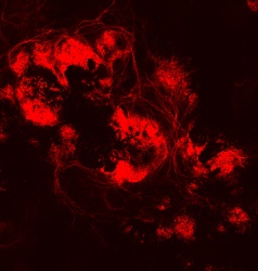
Image Source: Fluorescence microscopy: Human retinal ganglion cells at day 50.
Credit: Courtesy of Johns Hopkins Medicine; image provided by Valentin Sluch
“By the 30th day of culture, there were obvious clumps of fluorescent cells visible under the microscope,” says lead author Valentin Sluch, Ph.D., a former Johns Hopkins biochemistry, cellular and molecular biology student and now a postdoctoral scholar working at Novartis, a pharmaceutical company. Sluch completed this research at Johns Hopkins before transitioning to Novartis.
Work completed through the following,
Wilmer Eye Institute’s Glaucoma Center of Excellence and its Stem Cell Ocular Regenerative Medicine Center, whose co-director is Zack.